
Share
Follow Us
HEPATOLOGY, October 1998, p. 1154-1158, Vol. 28, No. 4
Clinical Challenge
Management of Ectopic Varices
Ian D. Norton1, James C. Andrews2, and Patrick S. Kamath1
From the 1 Division of Gastroenterology and Hepatology and the 2 Department of Diagnostic Radiology, Mayo Clinic and Foundation, Rochester, MN
INTRODUCTION
The term “ectopic varices” is sometimes reserved for abnormally dilated veins associated with gastrointestinal mucosa and, therefore, with the potential for gastrointestinal hemorrhage. However, the term has also been used loosely to describe portosystemic collateral veins in the abdominal wall and retroperitoneum. The distinction between “ectopic varices” and collaterals that are commonly found on the abdominal wall and retroperitoneum of patients with portal hypertension is one of semantics. Thus, ectopic varices may be best defined as large portosystemic venous collaterals occurring anywhere in the abdomen except in the cardioesophageal region.
Ectopic varices are an unusual cause of gastrointestinal hemorrhage, but account for up to 5% of all variceal bleeding.1 The clinician caring for patients with gastrointestinal bleeding must be aware of this entity, because diagnosis and management of ectopic varices differ from that of esophagogastric varices. Furthermore, the prognosis from bleeding ectopic varices may be poor, with one study quoting 40% mortality at initial bleed from duodenal varices.2 The literature on this subject consists mainly of small series and case reports with no randomized trials of therapeutic modalities. However, a review of the literature does provide sufficient information from which rational management decisions can be made.
PREVALENCE
Ectopic varices account for between 1% and 5% of all variceal bleeding. 1,3 Ectopic varices are a relatively common finding at endoscopy in patients with portal hypertension. The prevalence seems to be related to the cause of the portal hypertension and the technique used to show the varices. In patients with intrahepatic portal hypertension, duodenal varices are seen in 40% of patients undergoing angiography,3 whereas anorectal varices have been reported in between 10% and 40% of cirrhotic patients undergoing colonoscopy. 4,5 It is important to differentiate anal varices from hemorrhoids: Anal varices collapse with digital pressure, whereas hemorrhoids do not.4 In patients with portal hypertension caused by obstruction of the portal or splenic veins, duodenal varices are more prevalent than in patients with intrahepatic portal hypertension. The prevalence is higher if angiography is used to show varices. In fact, most patients with portal or splenic vein thrombosis are likely to have duodenal varices shown on angiography. 6,7 The majority of patients with duodenal varices visualized on endoscopy have extrahepatic portal hypertension. In contrast to duodenal varices, it appears that most cases of varices in other portions of the small intestine and colonic varices are seen in patients with intrahepatic portal hypertension who have previously undergone abdominal surgery.6 In the west, because the prevalence of extrahepatic portal hypertension is low, most bleeding from ectopic varices is usually associated with intrahepatic portal hypertension. 6,8 Stomal varices are a particularly common cause of ectopic varices and occur in patients with intrahepatic portal hypertension caused by primary sclerosing cholangitis.6
Although ectopic varices can occur at several sites, bleeding ectopic varices are most commonly found in the duodenum and at sites of previous bowel surgery including stomas. In a review of 169 cases of bleeding ectopic varices, 17% occurred in the duodenum, 17% in the jejunum or ileum, 14% in the colon, 8% in the rectum, and 9% in the peritoneum. In the review, 26% bled from peristomal varices and a few from infrequent sites such as the ovary and vagina.
ETIOPATHOGENESIS
Ectopic varices are natural portosystemic shunts, resulting from portal hypertension, and may occur at any site in the gut. Collaterals occur where the portal venous system is in apposition to systemic veins and are typically in the gastroesophageal junction and in the anorectum but can also occur around the umbilicus, ovaries, and bare area of the liver. Resistance within these collateral vessels is initially high; in contrast, resistance in the portal vasculature is low. Thus, blood flows preferentially through the portal venous circulation, and collateral vessels remain collapsed. However, with the development of portal hypertension, intrahepatic vascular resistance increases, allowing the portosystemic collaterals to open. In extrahepatic portal venous obstruction when the defined portosystemic collaterals are obstructed, less well-defined collaterals, such as in the duodenum and colon, open up. Surgery that results in apposition of bowel to abdominal wall or other structures drained by the systemic venous circulation also results in collaterals being formed in unusual sites. The best recognized of these ectopic varices is stomal varices formed at ileostomy sites in patients with primary sclerosing cholangitis who have undergone colectomy with creation of an ileostomy for inflammatory bowel disease, usually ulcerative colitis.9
Ectopic varices account for between 1% and 5% of all variceal bleeding. 1,3 Ectopic varices are a relatively common finding at endoscopy in patients with portal hypertension. The prevalence seems to be related to the cause of the portal hypertension and the technique used to show the varices. In patients with intrahepatic portal hypertension, duodenal varices are seen in 40% of patients undergoing angiography,3 whereas anorectal varices have been reported in between 10% and 40% of cirrhotic patients undergoing colonoscopy. 4,5 It is important to differentiate anal varices from hemorrhoids: Anal varices collapse with digital pressure, whereas hemorrhoids do not.4 In patients with portal hypertension caused by obstruction of the portal or splenic veins, duodenal varices are more prevalent than in patients with intrahepatic portal hypertension. The prevalence is higher if angiography is used to show varices. In fact, most patients with portal or splenic vein thrombosis are likely to have duodenal varices shown on angiography. 6,7 The majority of patients with duodenal varices visualized on endoscopy have extrahepatic portal hypertension. In contrast to duodenal varices, it appears that most cases of varices in other portions of the small intestine and colonic varices are seen in patients with intrahepatic portal hypertension who have previously undergone abdominal surgery.6 In the west, because the prevalence of extrahepatic portal hypertension is low, most bleeding from ectopic varices is usually associated with intrahepatic portal hypertension. 6,8 Stomal varices are a particularly common cause of ectopic varices and occur in patients with intrahepatic portal hypertension caused by primary sclerosing cholangitis.6
On rare occasions varices may be present in the colon in the absence of portal hypertension caused by anomalies of portal venous outflow, such as congenital anomalies of normal portosystemic anastomoses,10 abnormal vessel structure,11 or arteriovenous fistulae.12 Familial occurrence of colonic varices has also been described.11
Wall tension is thought to be the major determinant of hemorrhage from varices.13 Tension in the varix wall is proportional to transmural pressure across the vessel wall and the radius of the vessel. Therefore, the major determinants of rupture of ectopic varices, as in esophageal varices, are likely to be vessel size and portal pressure.
MANAGEMENT OF BLEEDING ECTOPIC VARICES
Because of the infrequency with which bleeding ectopic varices present, there have been no randomized trials on the management of this condition and it is unlikely that there ever will be such trials. Therefore, management depends on local expertise and the cause of portal hypertension. The management of varices in the abdominal wall and retroperitoneum (also called intra-abdominal varices), of stomal varices, and of varices around the biliary tree is different from that of other ectopic varices and is discussed separately.
Most patients with ectopic variceal hemorrhage present with sudden, profuse melena or hematochezia. Duodenal varices may also be associated with hematemesis. Bleeding from ectopic varices is suspected in all patients with portal hypertension and gastrointestinal bleeding in whom upper and lower gastrointestinal endoscopy have not shown a bleeding source.
Initial management involves clinical assessment and hemodynamic stabilization. The use of octreotide to reduce splanchnic blood flow and variceal pressure may be beneficial in patients bleeding from esophagogastric varices.14 The role of this drug in the control of bleeding from ectopic varices has not been addressed. It is our routine to commence an octreotide infusion (50 µg/h) in all patients presenting with suspected portal hypertension-related bleeding. It is unclear whether this intervention helps, and is only justified in light of the safety profile of octreotide. Similarly, terlipressin, which has been beneficial in the management of bleeding esophageal varices15 may be tried, but this drug is not currently available in the United States.
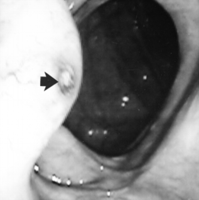
Because brisk upper gastrointestinal bleeding can result in hematochezia, all subjects with suspected variceal hemorrhage undergo emergency upper endoscopy as a first line investigation. If the upper endoscopy fails to show a source of upper gastrointestinal hemorrhage, colonoscopy is performed after a rapid colonic purge, typically 3 L of polyethylene glycol solution delivered via a nasogastric tube. In the article by Lebrec and Benhamou,6 22% of patients with ectopic varices bled from the colon or rectum. Colonic (Fig. 1) and rectal varices are identified as serpiginous vessels projecting into the lumen. If the colon appears normal and bleeding continues, angiography may be useful in identifying varices or in localizing a nonvariceal source of hemorrhage. Even in the face of massive bleeding, conventional transfemoral angiography may not show the site of variceal bleeding.
|
Fig. 1. Colonic varices as seen at colonoscopy. The bleeding site is shown (arrow). |
Most ectopic varices are within reach of standard endoscopy.6 Several studies have reported successful therapy of duodenal varices with sclerosant injection.16 Our experience with sclerosant has been less than satisfactory, especially with colonic varices. This may be because sclerosant may be diluted in large varices beyond a concentration adequate to obliterate the varix. Successful control of bleeding after the injection of cyanoacrylate or thrombin/sclerosant combination has also been reported.17 There are no data regarding band ligation of ectopic varices. It is our opinion that if the entire varix cannot be banded there is the risk of causing a wide defect in the varix after sloughing of the band, rendering the banding technique unsafe for large ectopic varices. We recommend banding only if the diameter of the varix is less than the diameter of the endoscope.
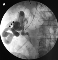
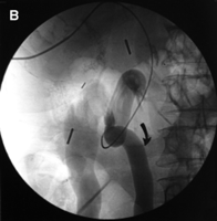
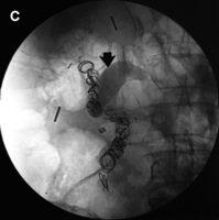
Extensive literature exists regarding the efficacy of embolization therapy using radiological techniques in the short-term therapy of bleeding esophagogastric varices. A number of techniques have been used to embolize the varices, including steel coils, thrombin, gelfoam, collagen, and autologous blood clot18 either alone or in combination with a variety of sclerosants. In the acute setting, these techniques control bleeding in up to 94% of cases. 19,20 Unfortunately, these techniques do not decompress the portal system, resulting in high rebleeding rates over 1 year.18 The experience with percutaneous embolotherapy for ectopic varices is limited to case reports.21-23 The transhepatic approach is used to catheterize the portal vein; the catheter is passed into the mesenteric veins, and angiography is directly performed. The transhepatic approach is preferred, especially in the patient with active bleeding, because access to the portal venous system is much quicker than when the transjugular approach is used. The ectopic varices and their drainage into the systemic venous system are identified (Fig. 2). Unlike arterial embolization for gastrointestinal hemorrhage, the goal in this setting is not to occlude the bleeding site itself, but to occlude the feeding vein (that is, the vein on the portal venous side) to the ectopic varices. This allows the varices to drain into the low pressure systemic veins, while protecting them from the high pressure portal system (Fig. 2). Steel coils are the preferred agent, because they provide a permanent, focal occlusion. They are available in a variety of sizes, which allows occlusion of large veins without difficulty. The transhepatic approach to variceal embolization may be difficult to apply in patients with portal vein occlusion. The transjugular approach may be used if a transjugular intrahepatic portosystemic shunt (TIPS) is planned immediately after embolization of the varix. We restrict this approach only to patients who are on a waiting list for liver transplantation.
|
Fig. 2. (A) Angiographic view of the colonic varices (arrowheads) shown in Fig. 1. The transhepatic catheter is in the feeding mesenteric vein branch. (B) A late image from the same series shows the large draining systemic vein. (C) Angiography shows stasis in the feeding vessels (arrow) and disappearance of the varices. |
If the patient continues to bleed in spite of embolization of the ectopic varices, options include either creating a TIPS or proceeding with surgery. At surgery, the varix is ligated and, in some circumstances, a portosystemic shunt is created. The choice of TIPS or surgery depends on the underlying liver function. Surgery is preferred in patients of Child-Pugh class A and in patients with an extrahepatic portal vein thrombosis as the cause of portal hypertension.
Several recent publications have emphasized the role of TIPS in the management of bleeding ectopic varices caused by intrahepatic portal hypertension.24-29 In the largest series to date, nine patients with intestinal varices received TIPS.26 Five of these patients were Child-Pugh class C at the time that the procedure was performed. Six were actively bleeding at the time of the procedure. Five of these patients stopped bleeding with the TIPS and one required embolization of the portal feeding vessel to stop bleeding. There was no procedural mortality and no recurrence of bleeding in the follow-up period (median, 7 months). However, two patients died within 5 days of the procedure from multisystem failure, and three more died within 6 months. This illustrates the generally bleak prognosis in patients of poor liver function who bleed from portal hypertension. Furthermore, up to 50% of TIPS will stenose by 6 months. It appears that in patients with intrahepatic portal hypertension, TIPS offers a highly effective means of controlling hemorrhage in the short term; if patients have good liver function and long-term survival is expected, a surgical portosystemic shunt may be preferred. It must be emphasized that only a nonselective portosystemic shunt, such as portocaval, mesocaval, or central splenorenal shunt, will adequately decompress ectopic varices. In case of extensive thrombosis of the portal venous system, nonconventional portosystemic shunts using large collateral vessels may be performed.30
There are no data available regarding the use of adrenergic![]() -blockers or nitrates in the long-term management of patients with ectopic varices. A meta-analysis of randomized, controlled trials comparing a nonselective
-blockers or nitrates in the long-term management of patients with ectopic varices. A meta-analysis of randomized, controlled trials comparing a nonselective ![]() -blockade versus placebo showed the efficacy of
-blockade versus placebo showed the efficacy of ![]() -blocker therapy for secondary prophylaxis of bleeding from esophagogastric varices.31 Therefore, it seems logical that these modalities could be tried in patients with ectopic varices. However, in one study,
-blocker therapy for secondary prophylaxis of bleeding from esophagogastric varices.31 Therefore, it seems logical that these modalities could be tried in patients with ectopic varices. However, in one study, ![]() -blocker therapy did not appear to be effective in the management of peristomal varices.9 We recommend the use of
-blocker therapy did not appear to be effective in the management of peristomal varices.9 We recommend the use of ![]() -blocking agents in the prevention of rebleeding or when patients cannot undergo a surgical shunt. This is usually when portal venous thrombosis is so extensive that a suitable vein (8 mm or more in diameter) cannot be found for anastomosis with a systemic vein.
-blocking agents in the prevention of rebleeding or when patients cannot undergo a surgical shunt. This is usually when portal venous thrombosis is so extensive that a suitable vein (8 mm or more in diameter) cannot be found for anastomosis with a systemic vein.
Intra-abdominal Varices. Veins in the abdominal wall may rupture externally. More often, veins around the falciform ligament of the liver rupture into the peritoneal cavity. Fewer than 20 such cases have been reported in the literature. External rupture is easily recognized and treated with local compression and surgical ligation. Rupture of intra-abdominal varices is characterized by a rapid increase in ascites, particularly in the presence of abdominal pain, and is associated with a decrease in the hematocrit. The diagnosis is confirmed by a heavily blood-stained ascitic fluid tap. The diagnosis is further suggested by computed tomography scan demonstration of retroperitoneal varices and intra-abdominal hemorrhage.
Management of this situation is made more difficult by the relative contraindication to transhepatic obliteration of the varices caused by the accompanying ascites. The transjugular, intrahepatic approach to the portal system is another possibility for angiographic embolization therapy. In appropriate subjects, TIPS placement at the time of variceal obliteration may be considered, but in an acutely ill patient it should probably be deferred unless the initial embolization therapy fails to arrest hemorrhage. Emergency surgery may be performed in these patients with the aim of ligating the bleeding varix, but this carries a high mortality.32 Because such patients are usually quite ill at the time of surgery, no attempt is made to create a portosystemic shunt.
Stomal Varices. The most common scenario for stomal varices is in patients with ileostomies after proctocolectomy for inflammatory bowel disease with associated primary sclerosing cholangitis.9 Stomal varices are recognized by the purplish hue around the stoma. The patient is usually aware of the bleed early in its clinical course and may be taught how to control the hemorrhage with pressure. Probably because the patient can control hemorrhage easily, the mortality from a stomal variceal bleed is relatively low (3%-4%).27 Thus, a relatively conservative approach to stomal varices is recommended.
Simple local measures, such as pressure dressings, epinephrine-soaked gauze, gel foam, and suture ligation, have all been used with success for the initial bleeding episode.27 Injection sclerotherapy has also been used, but this may result in mucosal ulceration, peristomal skin necrosis, and stricturing of the stomal orifice.33 Mucocutaneous disconnection of the stoma with ligation of the portosystemic channels has been performed under local anesthesia and is a less invasive therapy than refashioning the entire stoma, but long-term results with both procedures are usually suboptimal. We do not recommend either of these procedures as effective long-term treatment of stomal varices. In patients with uncontrolled or recurrent bleeding we advocate transhepatic obliteration of the bleeding stomal varices. In some patients this is insufficient to prevent further bleeding and a portosystemic procedure should be contemplated. Because many patients with stomal varices are candidates for liver transplantation, a TIPS procedure is preferred to a surgical portosystemic shunt.
Biliary Varices. In extrahepatic portal vein thrombosis, varices may occur around the gallbladder in 30% of patients.34 They are usually asymptomatic and require no therapy. Varices can also occur in portal venous obstruction around the common bile duct resulting in extrahepatic biliary obstruction, cholangitis, and hemobilia. The appearances of a biliary tree on endoscopic retrograde cholangiopancreatography may mimic that of primary sclerosing cholangitis.35 Optimal therapy requires both a surgical portosystemic shunt as well as a biliary drainage procedure, which may be either surgical, endoscopic, or radiological.
SUMMARY
The management of ectopic varices is difficult and must be performed by a multidisciplinary team approach of hepatologists, endoscopists, interventional radiologists, and surgeons. Although there are no large series on this subject, based on our experience and the literature of case reports, small series, and reviews, we present this systematic approach for any patient presenting with bleeding from possible ectopic varices (see Fig. 3).
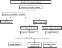
|
Fig. 3. An algorithmic approach to the patient with bleeding ectopic varices. |
Footnotes
Abbreviations: TIPS, transjugular intrahepatic portosystemic shunt.
Address reprint requests to Patrick S. Kamath, MD, Division of Gastroenterology and Hepatology, Mayo Clinic, 200 First Street SW, Rochester, MN 55905. Fax: (507) 284-0538; e-mail: kamath.patrick@mayo.edu.
Received August 3, 1998; accepted August 10, 1998.
Copyright © 1998 by the American Association for the Study of Liver Diseases.


