
Portal Hypertension: Prognosis and Treatment
Share
Follow Us
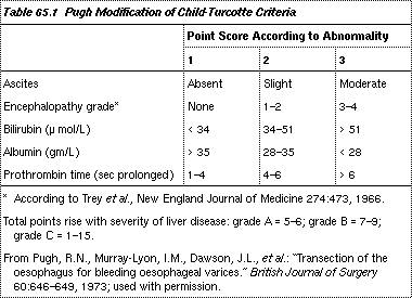
Prognosis is critically dependent on liver function. Liver failure is often the prime cause or a significant associated factor in mortality. The in-hospital (all inclusive) mortality rates with GI bleeding in Child-Pugh grade A (5 to 10%), B (15 to 25%), and C (50 to 70%) patients allow stratification of patients to improve assessment of outcome.
Treatment:
All patients with portal hypertension who have GI hemorrhage must be hospitalized. About 50 to 75% of bleeding episodes in cirrhotic patients are a direct consequence of portal hypertension: Bleeding is from either ruptured gastroesophageal varices or diffuse oozing from the gastric mucosa (congestive gastropathy). In congestive gastropathy, mesenteric venous hypertension leads to congestion of the gastric mucosa, particularly the fundus, rendering it more susceptible to damage and bleeding even with minor insults (eg, modest alcohol or aspirin ingestion). Other causes of GI bleeding (eg, duodenal or gastric ulcer, Mallory-Weiss lacerations) account for bleeding in the remaining patients.
Standard resuscitation measures include fluid and blood transfusions. Sedatives should be avoided, in view of possible encephalopathy. In the latter, cleansing enemas will remove blood products from the bowel, and lactulose will reduce hepatic encephalopathy. To determine the site of bleeding, endoscopy within 24 h of admission is advisable.
The most difficult, lethal bleeding is caused by variceal rupture. Emergency measures to stop variceal bleeding include mechanical balloon tamponade, vasoconstrictor drugs, and particularly endoscopic sclerotherapy, which is considered the first choice of therapy. Surgery should be avoided, if possible, because of the high operative mortality rate.
Types of balloon devices for esophageal tamponade are the Minnesota tube with an esophageal suction port, the Sengstaken-Blakemore tube with both an esophageal and gastric balloon, and the Linton-Nachlas tube with only a large gastric balloon. In practice, all are equally effective but potentially dangerous. Only experienced physicians should use these devices; the complication rate is 10 to 20%, even in experienced hands. Complications include aspiration pneumonia, esophageal rupture, and asphyxia.
Vasoactive agents that lower portal pressure are vasopressin (20 u. IV over 10 to 20 min) and a long-acting analog (terlipressin, and somatostatin or one of its analogs). Prolonged vasopressin infusions at lower dosage (eg, 0.1 to 0.4 u./min given over > 4 h) should be avoided due to side effects of coronary and mesenteric vasoconstriction that can lead to infarction, renal shutdown with oliguria, and local tissue necrosis if the IV infusion extravasates. Besides, long-term use is limited by the development of tachyphylaxis. Concomitant sublingual nitroglycerin (0.3 mg q 1 h) may help avoid some of the side effects of vasopressin. European experience with terlipressin suggests that its efficacy and side effects are comparable to vasopressin. Somatostatin may prove to have fewer side effects and equivalent efficacy.
The procedure of choice is endoscopic sclerotherapy. Variceal injections of several types of sclerosing agents effectively control acute bleeding and, later as elective therapy, eradicate varices once patients have stabilized. Complications of sclerotherapy include esophageal ulceration and perforation and, rarely, pulmonary embarrassment or portal vein thrombosis.
Surgery has a limited role. If bleeding is unresponsive or recurrent, the simplest, safest intervention is esophageal transection with a stapling gun. This controls bleeding in 90 to 95% of cases. A beneficial effect on survival has yet to be shown with any form of surgical therapy.
Chronic therapy:
Once the acute bleeding episode is over and the cirrhotic patient becomes stable , further treatment may be with drugs, sclerotherapy, or surgery. Propranolol appears to reduce rebleeding risk in a minority of patients, but responders cannot be easily identified, so routine use cannot yet be recommended. Other drugs (eg, clonidine, prazosin, ketanserin, and verapamil) are even more experimental.
Sclerotherapy is currently the procedure of choice, but changes in long-term prognosis remain unproven. For patients who continue to rebleed despite sclerotherapy, surgical options include liver transplantation, if indicated, or a shunt procedure to decompress the portal venous system into the systemic circulation. Various types of portacaval and mesocaval shunts, as well as the splenorenal shunts (including the distal splenorenal shunt, the Warren shunt), all divert some or all of the portal blood away from the liver. These procedures tend to precipitate hepatic encephalopathy, a disabling condition that has limited the value of portal decompression. Other side effects include deterioration of liver function and onset or progression of hepatic iron deposition (hemosiderosis).


