Share
Follow Us
Histopathology of the Liver in Children With Chronic Hepatitis C Viral Infection
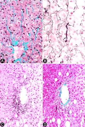
Fig. 4. (A) Pericentral pericellular fibrosis characterized by wire-like bands of collagen radiating from the terminal hepatic venule along the space of Disse (Masson trichrome stain). (B) Reticulin stain highlights a dense band of mature collagen filling up the space of Disse (between arrows) and compressing adjacent hepatocytes. (C) Hematoxylin-eosin stain demonstrating centrilobular necroinflammatory activity and endothelialitis (immunohistochemical stain for cytokeratins [monoclonal antibody clone AE1] specific for bile duct epithelium was negative in this area). (D) Trichrome stain of the same field shown in (C) demonstrates delicate bands of collagen extending from the venule into the lobule along the space of Disse.